Positron Emission Tomography (PET) is a nuclear medicine scan that uses a radioactive sugar solution to create images of how your organs and tissues function, and to identify normal vs. abnormal (cancer) cells. When the sugar solution concentrates in certain areas of the body, the PET scan picks-up the pattern of radioactivity and creates 3-dimensional images. Different colours and degrees of brightness in a PET scan image represent different levels of organ and tissue function. Cancer cells require more sugar to function, and therefore, appear brighter in a PET scan image.
To improve the accuracy of the diagnosis, we have integrated a CT (Computed Tomography) scanner. The result is a fusion of two sets of images: One that depicts the functional images of abnormal cells (PET) and the other demonstrates the structure of the tissues and organs (CT). The combination of these images will improve the physician’s ability to determine exactly where in the body the changes are taking place.
BEFORE THE TEST
- The evening before your scan, do not consume sugary drinks or foods, such as juice, soda, rice, pasta, white bread or baked goods. For the full day prior to your scan, choose higher protein foods, such as nuts, seeds, meat, poultry, fish, eggs, cheese, beans and vegetables.
- Discontinue vitamins, supplements and caffeine the evening before your scan. You may take your regular medications, unless they contain caffeine.
- Do not exercise for 12 hours prior to your scan and minimize your physical activity for the full day prior to your scan.
- Do not eat or drink anything other than plain water for 6 hours prior to your scan.
- Wear loose, comfortable layers of clothing that are free of metal buttons and zippers.
DURING THE TEST
- You will be given a consent form and questionnaire to complete.
- We will measure your blood sugar level using a glucometer.
- A radioactive sugar solution called FDG (18-Fluoro-deoxyglucose) will be injected intravenously into your bloodstream, and you will rest for 45 minutes to allow the solution to distribute throughout your body.
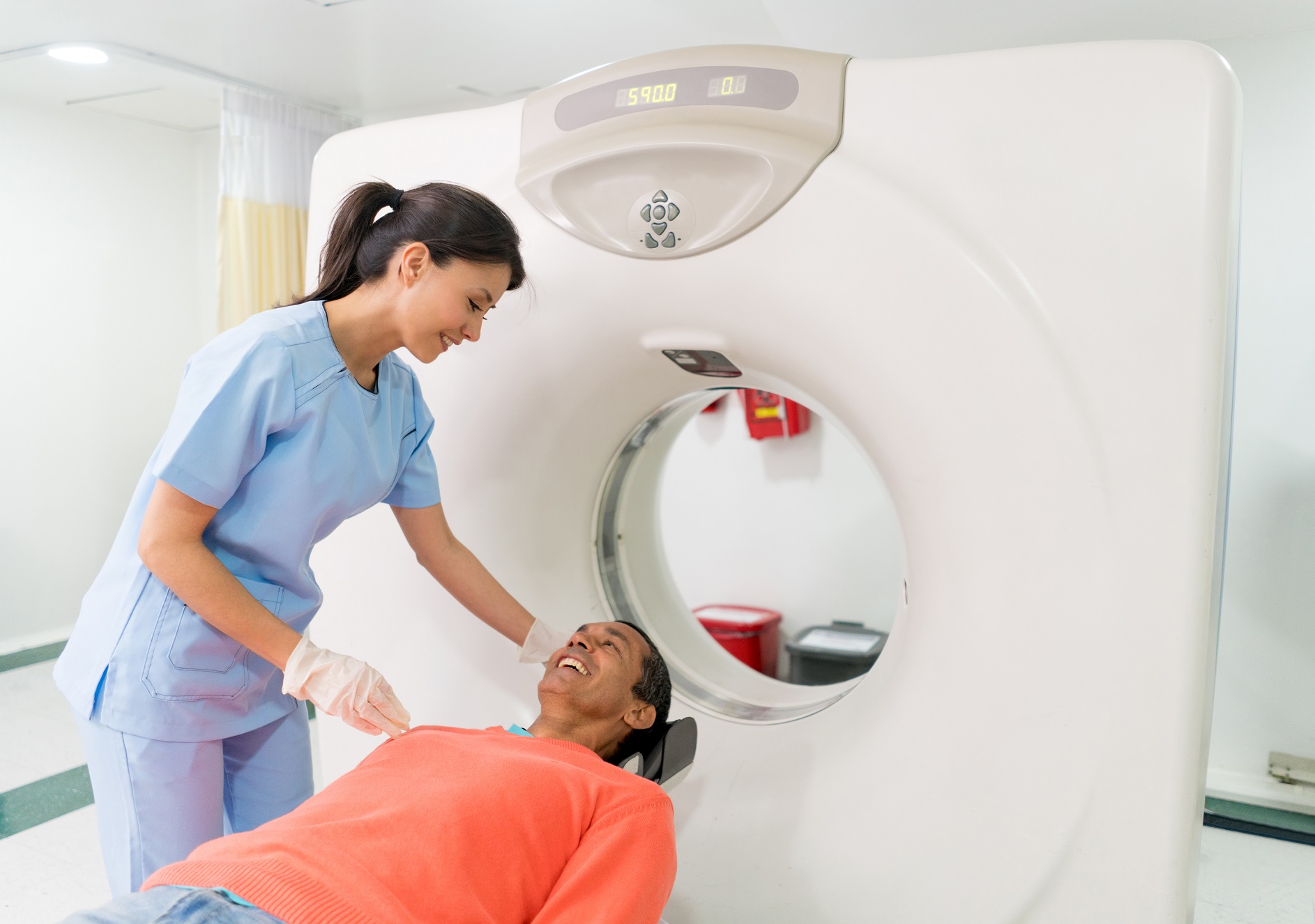
- You will be asked to lie as still as you can during the scan.
- The imaging process takes approximately 30 minutes, and you can communicate with your technologist throughout the scan.
Locations
Google Reviews
They run right on time and the technician was very informative with step-by-step details as to what was happening. He was also very sympathetic of my phobia and helped me through it.
– LYNDA B.