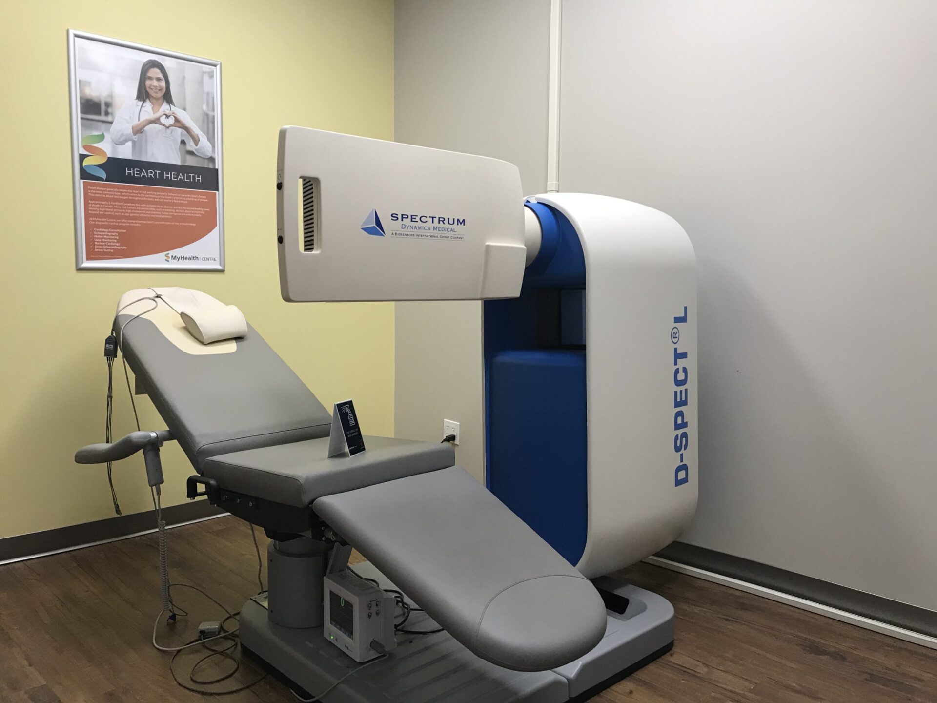
DIAGNOSTIC TESTS
Nuclear Cardiology (MPI)
A test to determine how well your arteries are delivering blood to your heart.
Myocardial Perfusion Imaging (MPI), also known as a Nuclear Stress Test, is done to determine how well the coronary arteries are delivering blood to your heart, as well as areas of the heart muscle that aren’t getting enough blood flow.
If the test shows a lack of blood flow during exercise or stress, but is normal at rest, it could mean that an artery that carries blood to your heart is narrowed or blocked. If the test shows a lack of blood flow to a portion of the heart muscle during exercise or stress and at rest, it could mean that your heart muscle is scarred, possibly from a past heart attack.
MPI is useful in patients with chest discomfort to see if the discomfort comes from a lack of blood flow to the heart muscle caused by narrowed or blocked heart arteries (angina). Myocardial perfusion imaging doesn’t show the heart arteries themselves, but it can tell your doctor with good certainty if any heart arteries are blocked and how many.
BEFORE THE TEST
- You should be fasting (except water) for 4 hours before the test
- Insulin-dependent diabetics should take their insulin with a light meal 2 hours before the test.
- Discontinue all caffeine products 24 hours before the test. This includes all tea, coffee, decaffeinated tea/coffee, pop, chocolate, Tylenol 2 & 3 and/or medications containing caffeine.
- Wear loose fitting clothing, such a t-shirt, track pants, athletic shoes, etc.
- Bring a list of all current prescription medications and check with your physician regarding the discontinuation of any heart medications, such as Beta-Blockers like Metroprolol or Atenolol, as well as Calcium Channel Blockers like Diltiazem or Verapamil.
- Do not take erectile dysfunction medications, such as Viagra, Cialis, Levitra, etc. for 48 hours before the test.
- If you are pregnant or there is a possibility of pregnancy, or if you are breastfeeding, Nuclear Medicine Stress Testing may not be appropriate for you at this time. Please consult with your physician.
DURING THE TEST
The test will take place in two parts. These parts may be completed in one or two days.
Rest Study: 1.5-2 Hours
- For this portion of the test the technologist will administer via intravenous injection a small amount of radioactive tracer, which is carried by the blood stream to your heart. There are no side effects with this injection. Approximately 15-45 minutes after your injection, the technologist will take pictures of your heart for approximately 20 minutes with a special gamma camera that detects the injection. For the imaging, you may be sitting in a specially designed imaging chair or lying on a bed and while the pictures are being taken, it’s important to remain very still during this time to avoid blurring the images.
2. Stress Study: 2-2.5 Hours
- For this portion of the test, ECG electrodes will be place on your chest to monitor your heart rhythm throughout the test. An intravenous line will also be placed in the arm or hand. You will begin to exercise on a treadmill, during which time your heart rate and blood pressure will rise. These are normal responses that are being closely monitored with your ECG.
- If you are unable to exercise adequately on a treadmill, similar results can be achieved using a drug called Persantine. Persantine mimics the effects of exercise by dilating the blood vessels of the heart, allowing for increased blood flow.
- Once you have reached your maximum level of exercise, the technologist will administer a small amount of radioactive tracer and you will continue exercising for an additional 1-2 minutes. If blood flow to the heart is limited due to coronary artery disease, then the amount of tracer in your heart is reduced. Following the Stress Test, the intravenous will be removed.
- It is very important that a set of pictures is taken after the stress test to compare to the resting images and assess the amount of blood supply to the heart at rest and during stress (exercise). Once the stress test is complete you will wait approximately 15-45 minutes afterwards before the imaging can begin. The wait time may vary based on individual characteristics to best optimize image quality, the technologist will advise how long you will be waiting before images.
AFTER THE TEST
You will be able to return to your normal activities when the test is complete, unless your physician or technician tells you otherwise. You can drive after the test. If you are travelling by plane, train or crossing the border within one week after your test, please inform the technologist.
Source: heart.org
Locations
- Brampton Chrysler
- Lindsay
- London Fanshawe
- Milton Cardiology
- Mississauga Cardiology
- Newmarket Cardiology
- Orangeville
- Oshawa
- Sarnia
- Scarborough
- Simcoe
- Sudbury Larch Imaging
- Toronto Davisville
- Toronto King
- Whitby
Google Reviews
This is an excellent place to get answers to health conditions. I would recommend them to family and friends. The staff are friendly and professional.
– JUSTIN P.

